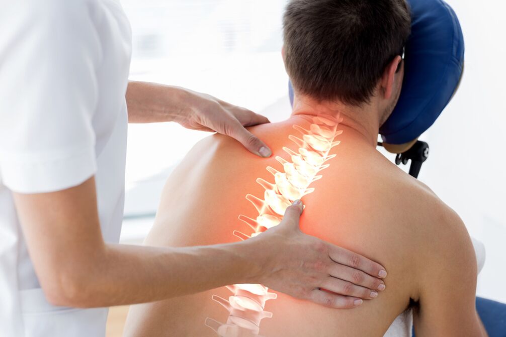
Osteochondrosis - dystrophic changes in the spine associated with age-related tissue aging. 80% of the pathology is associated with genetic information, the rest is the influence of external factors.
Osteochondrosis- mainly contributes to the development of human disease:
- Increased life. Over time, metabolism slows down, tissue nutrition is disrupted, and destructive regulatory systems begin to dominate constructively.
- Walk upright. Standing on his feet, the man received an unequal load on different parts of the spine, was able to move more - to twist, to stretch. Abnormal lateral flexion - scoliosis - showed an uneven load on the muscles and small joints of the spine. This even increased the likelihood of developing the disease in the department that protects the vertebrae of low mobility and rib cage - thoracic osteochondrosis.
- Acceleration. Rapid growth makes bones, muscles and cartilage more sensitive. The number and distribution of blood vessels are not sufficient to provide them with oxygen and essential nutrients.
- Lack of adequate physical activity. There are two extremes - sedentary work and only driving, or excessive stress in the gym, when the discs and cartilage wear out at an accelerated rate.
- Malnutrition. The predominance of fast carbohydrates, lack of protein, the use of carbonated beverages leads to a lack of high-quality building materials to maintain the health of the body's tissues.
- To smoke. Causes long-term vasospasm - tissue malnutrition, acceleration of degenerative processes
- Urbanization, a large number of traumatic objects in the environment cause damage to the spine, secondary osteochondrosis.
Types of osteochondrosis
Due to localization
- Osteochondrosis of the cervical spine
- Injury of the thoracic spine
- Lumbar osteochondrosis
- General osteochondrosis - neck and spine, thoracolumbar, lumbosacral and other joints
The most common changes in the most mobile parts are the cervical and lumbar. The painful place is the transition of the mobile lumbar region to the stable sacral region.
By stage
- Primary - small changes in the center of the disc, compression of the nucleus, the appearance of cartilage cracks
- Progression of the disease - the cracks deepen, the height of the disc decreases, the diameter of the intervertebral foramen decreases. Compression of the roots of the spinal nerve causes pain and muscle spasms. Osteochondrosis of the spine is not only manifested by changes in the discs - due to the violation of the ratio of the vertebrae to each other, the cartilage on the surfaces of small joints is unevenly removed, arthrosis and arthritis develop.
- Complex osteochondrosis - symptoms: further degeneration of cartilage occurs - fractures of the cartilaginous ring connecting the trunks of two adjacent vertebrae are visible. Part of the nucleus comes out of nowhere and squeezes the roots, forming a spinal cord - disc herniation. A more serious problem is the separation of the collapsed part - sequestration hernia. Severe pain in the area responsible for the compressed nerve is disturbed by impaired sensitivity and movement.
- The body responds to increased load and excess mobility with the growth of bone tissue - osteophytes appear. They stabilize the spine, but reduce the range of motion. Bone hooks irritate muscle receptors and press on nearby vessels. With cervical osteochondrosis, it causes "vertebral artery" symptoms - dizziness, tinnitus, tremors in front of the eyes
Osteochondrosis of the cervical spine
With the advent of mobile phones and computerscervical osteochondrosiseven in adolescents: a prolonged unnatural position of the head with muscle tension overloads the vertebrae, their discs and joints.
Cervical osteochondrosis - symptoms
- Neck pain extending to the back of the head, upper back
- Sometimes headaches associated with cervical osteochondrosis mimic migraines - one-sided symptoms, intolerance to sounds and bright light, strong pulsation in the temples, bright flashes in front of the eyes.
- Frequent headaches that do not respond well to traditional tablets
- Constant pressure drops on antihypertensive drugs
- Dizziness and darkening of the eyes with sudden dizziness
- Especially after sleep, numbness in the fingers, a feeling of crawling on the skin
- Restriction of movement in the neck, crunching when trying to move. Patients have to turn their whole bodies to see something behind them
- Upper body sweating
- Tense muscles of the neck and shoulder girdle can be detected by palpation.
If determinedcervical osteochondrosis, treatment in the early stages prevents severe complications - compression of the vertebral artery with oxygen starvation of the brain, compression of the spinal cord.
Manifestations of osteochondrosis of the thoracic spine
Changes in the thoracic region are less developed, the motivating factors - back injuries, scoliosis, previous diseases of the spine (tuberculosis, nonspecific spondylitis, hemangiomas).
Symptoms of thoracic injury:
- Low back pain - aching, pulling, worsening after standing for a long time or sitting in an uncomfortable position. However, other possible causes with persistent pain complaints should be ruled out - pneumonia, pleurisy, tumors, intercostal neuralgias of a different nature, herpes zoster before the appearance of bubbles.
- Difficulty breathing, shortness of breath, inability to breathe deeply
- Thoracic osteochondrosis sometimes mimics attacks of angina pectoris - a person is treated by a cardiologist for a long time and the problem is in the patient's intervertebral disc.
Lumbar and lumbosacral osteochondrosis
In the structure of all types of osteochondrosis, these departments are reliable leaders, accounting for more than half of all diagnoses. This is because the greatest load falls on this area of the body, both on the feet and while sitting. If the body weight is not lifted properly, the load is in a bent position for a long time - the pulp nucleus of the intervertebral discs is compressed, the cartilage plates are compressed into the spinal cord - Schmorl's hernias are formed. . Excessive tension and muscle spasms disrupt the relative position of the small joints of the vertebrae - the joint cartilage is removed, mobility is reduced.
Several bad circles develop at once: muscle spasm causes pain - pain reflexively increases the contraction of muscle fibers, acute pain forces a person to restrict movement, protects the damaged area - decreases the strength of the muscle frame and spinal support, which increases its strength. instability, lumbar osteochondrosis progresses.
At the mobile access pointwaist waistwhen entering a stationary sacrum connected to a single monolith, there is a danger of the fifth vertebra slipping off the surface of the sacrum. This compresses the nerve endings, and radicular syndrome develops.
Symptoms of lumbar osteochondrosis
- Back pain, especially when sitting and standing. After rest, the horizontal position improves. With a long-lasting course, the pain is drawn to the usual, aching
- A sharp sudden lumbago when changing the position of the body, lifting weights, lifting heavy loads. The patient is stuck in an attack, it is difficult to correct, to start moving. Lumbago is usually associated with compression of the spinal cord, which develops sharply
- Transfer of pain to the gluteal region, legs. The largest nerve in the body, the sciatic, is a direct extension of the spinal cord, so patients with lumbar osteochondrosis are often concerned about sciatica.
- Because nerve fibers control the tone of muscles and blood vessels and regulate tissue nutrition, changes are noted in that part of the body where the patient's nerve is responsible. The limb feels colder than healthy. With long-term disease, muscle atrophy, dry skin and swelling are observed. Local immunity is reduced - any scratches, cuts, abrasions easily become a gateway for infection.
- Defeat of sensitive fibers leads to a violation of sensitivity - superficial and deep. The patient may be burnt or frozen because he does not feel a dangerous change in temperature.
- Very frightening symptoms - numbness of the skin of the perineum, loss of control over the pelvic organs. The patient does not feel the bladder is full, does not feel the need to empty the bowels. Over time, urine and feces begin to be expelled spontaneously, making it impossible to store them. In this case, the treatment of osteochondrosis of the spine and its complications is urgent surgery.
Diagnosis of osteochondrosis
It is performed by a neurologist or orthopedist after the therapist has ruled out pathology of the internal organs.
- The specialist studies the main complaints, their time of onset, development, the effect of drugs on the intensity of pain, rest, changes in rhythm of life.
- When the patient takes off his underwear, a mandatory external examination is performed - it is necessary to compare the condition and color of the skin in symmetrical parts of the body, tissue tone, reaction to various stimuli: pain, touch, cold. or heat. Signs of tension are identified, which indicate muscle tension and irritation of their tendons and integumentary membranes - fascia
- The neurological hammer will reveal the unity and symmetry of the reflexes
- The neurologist notes the volume of active (independent) and passive (performed by a doctor) movements in the joints, the ability to turn the head and upper body without involving the lower parts of the spine.
If necessary, send for additional examination
- Thermal imaging diagnostics
- ENMG (Electromyography): Radiography. In order to obtain the necessary information, it is carried out in at least two projections - directly and laterally. The image will provide information about the condition of the bone tissue, the severity of osteoporosis, the size and safety of the vertebral bodies, and detect osteophytes. Damaged discs are defined by the width and uniformity of the intervertebral fractures. The unevenness of the lower or upper border of the body will suspect a Schmorl hernia. Computed tomography is recommended to clarify the nature of changes in the bone structure of the spine. Multispiral examination allows three-dimensional modeling of vertebrae. If necessary, to study the condition of soft tissues - muscles, ligaments, intervertebral disc, MRI is prescribed.
It should be noted that the results of the study should be compared with the complaints and changes identified during the examination. The undetected detection of symptoms of spinal osteochondrosis and even a herniated disc does not require any serious action.
Treatment of osteochondrosis of the spine
Elimination of acute manifestations of the disease
- Severe pain and sharp muscle tension reinforce each other, preventing the inflammation from decreasing. Therefore, the first is to eliminate the pain.
- Prescribe injections of non-steroidal anti-inflammatory drugs, muscle relaxants - muscle relaxants.
- If these measures are not enough, blockade with painkillers and hormonal drugs
Radiofrequency denervation
A few days of bed rest is recommended
Once the symptoms have resolved, you should begin to move, gradually increasing your range of motion and load. In this case, active kneading and massage are undesirable due to possible complications.
Osteochondrosis: treatment without exacerbation
When the patient's condition stabilizes, the usual lethargy remainsosteochondrosis, treatment consists of several components:
- Medication. The same anti-inflammatory painkillers in tablets, capsules and ointments. A particular drug is selected by the doctor based on the patient's condition, lifestyle, concomitant diseases, the predominance of one or another component of osteochondrosis. The course of B vitamins will improve the conduction of impulses along the nerve, normalize tissue nutrition. The use of muscle relaxants will continue while maintaining increased muscle tone. There is no magic pill, no injection that can restore the vertebrae and cartilage to their original state. Medications relieve symptoms, improve mobility and performance. However, they cannot completely stop the disease.
- Physiotherapy. It is used for direct delivery of drugs to the painful area (electrophoresis), heating (paraffin, infrared radiation). Exposure to therapeutic currents relaxes muscles and improves the activity of nerve fibers. After a few sessions, the pain decreases and mobility is restored. Not prescribed for active inflammation
- Manual manipulation, massage, acupuncture, acupressure. Eliminate spasms by stretching and relaxing muscles. If only the upper layer of muscles is affected during the massage, manual therapy penetrates deeper, so the requirements for specialists are higher. Be sure to take an MRI first to learn the characteristics of a particular patient's anatomy
- Spinal traction. The vertebrae move away from each other, the normal distance between them is restored, and nerve compression is reduced. The procedure has contraindications, so only a doctor can prescribe it
- Physiotherapy. The most effective treatment. The only caveat is that it should be applied for life. Advantages - provides activity, improves mood, increases tissue tone. The best methods are a set of exercises recommended by your doctor, initial yoga asanas, Pilates, swimming. They are performed smoothly, without sudden and traumatic movements, stretch the tissue, the amplitude gradually increases.
- Get rid of bad nutrition and bad habits
- Adequate nutrition of tissues, good condition of blood vessels and adequate blood supply to the spine and surrounding structures are measures to prevent the development of osteochondrosis. Proper nutrition normalizes weight, reduces stress on the spine
Surgical treatment of osteochondrosis of the spine.Modern clinics have a large arsenal of minimally invasive interventions:
- Treatment and diagnostic blockade
- Radio frequency facet ablation
- Cold plasma and laser nucleoplasty
- Endoscopic removal of a herniated disc
- Microdiscectomy
Radiofrequency thermal ablation of facet joints
Special needles are placed exactly on the side of the intervertebral joints where the median branch of the Lyushka nerve passes. Electrodes are installed on the needles, the tip is heated to 80 degrees for 90 seconds. This causes the nerve to clot. The pain goes away.
Cold plasma nucleoplasty
A special cold plasma electrode is injected into the disc tissue through a needle inserted into the disc. Intradiscal pressure decreases, the hernia (bulge) is pulled in.
Microdiscectomy
Nerve roots and blood vessels adjacent to the herniated disc are compressed, there is extreme pain and various disorders of the innervation of the extremities. If the effects of conservative treatment are no longer effective, surgery to remove a herniated disc is the only possible solution for many patients. The operation is performed under anesthesia with a 2-3 cm incision using microsurgical equipment and tools. The operation takes 45-60 minutes. Pain syndrome is significantly reduced or completely eliminated in 95% of patients immediately after surgery. The next day the patient is allowed to walk and is soon discharged from the clinic.
Endoscopic removal of herniated discs:
A hernia or free sequestration is removed from the lateral intervertebral foramen. A 5 mm incision is made in the skin to place the tube. Muscles, fascia and ligaments are not damaged, they are pushed together using a system of tube retractors with a gradual increase in diameter. The operation is almost bloodless and lasts only 40-50 minutes. Patients can return to normal after three weeks. The risk of complications is minimal.
In case of complications, decompression and stabilization operations are performed during a large disc herniation, severe compression of the spinal nerve root and spinal cord. If there are signs of sudden loss of sensitivity, movement, pelvic dysfunction, the patient should be taken to a neurosurgeon immediately. The sooner it is possible to overcome the depression, the more complete the recovery will be, and the sooner the person will return to normal life. In this case, surgical treatment is aimed at decompression of compressed nerve structures and stabilization of the affected segment. This is a hemi or laminectomy. Fixation is performed by a transpedicular system, together with an inter-body cage that provides 360-degree fusion. Interspinous stabilization of the spine is widely used. Today there are several interspinous implants. Microdiscectomy combined with interspinous stabilization, especially in the elderly, can significantly increase the effectiveness of long-term outcomes and reduce the likelihood of recurrent disc herniation.


